

TOOTH HAMARTOMA
GEORGE JISS MARY, KUNJMON RENJU M, JAMES JESLIN
Department of Oral Medicine and Radiology, Annoor Dental College and Hospital, Muvattupuzha, Kerala
Received: 17 – 08 – 2022
Revised: 20 – 08 – 2022
Accepted: 26 – 08 -2022
Address for correspondence: Dr Jiss Mary G, Department of Oral Medicine and Radiology, Annoor Dental College & Hospital, Muvattupuzha, Email ID: jissnoel@gmail.com
This is an open access journal, and articles are distributed under the terms of the Creative Commons Attribution-Noncommercial ShareAlike 4.0 license, which allows others to remix, tweak, and build upon the work non-commercially, as long as appropriate credit is given and the new creations are licensed under the identical terms
How to cite this article: George J M, Kunjmon R M, James J. Tooth Hamartoma. J Oral Biomed Sci 2022; 1: 101-4
Key words: Compound Odontome, Hamartoma
A 21 year old male came to the department of oral medicine and radiology with chief complaint of slow growing swelling on left lower jaw since 2yrs. Patient was asymptomatic except for mild swelling on lower left side of face which became apparent only since last 6 months.
On inspection – Mild facial swelling noted on left side; extended from canine to first molar region. On palpation – swelling was painless, hard and non-mobile – with obliteration of vestibule
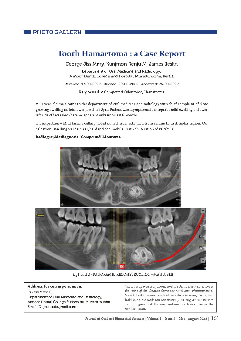
Radiographic diagnosis – Compound Odontome
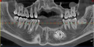

Fig1 and 2 – PANORAMIC RECONSTRUCTION –MANDIBLE

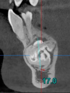
Fig 3 and 4 – Conglomerrate masses of opacifications noted in the lower left mandible with well formed enamel, dentin and pulp space with enlarged follicular space, Maximum dimension Bucco- lingually – 17.6mm, Anteroposteriorly – 20.4mm, Superoinferiorly – 17.0mm
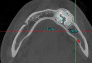
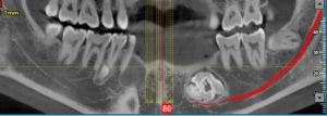
Fig 5 and 6 – Thinning of buccal cortical plate with buccal expansion, thinning of lingual cortical plate, compression of inferior alveolar canal
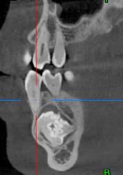
Fig 7 – Close proximity of opacification to the periapex of 34, 35 with no root resorption, displacement noted for 34, 35

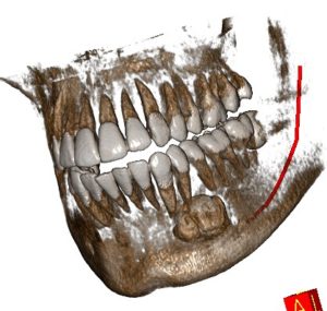
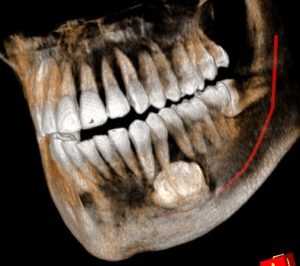
Fig 8 – 3D Reconstructed Image
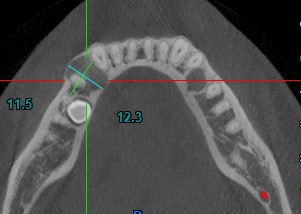
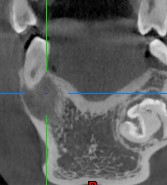
Fig 9 – Well defined hypodense region noted distal to 43 of size 11.5 mm X 12.3 mm involving periapical aspect of 44 with corticated border and mild scalloping, buccal cortical plate expansion and perforation noted, Lingual cortical plate thinning noted, Supernumerary tooth noted apical to 45 with no evident root formation.
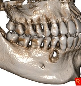
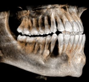
Fig 10 – 3D Reconstructed Image